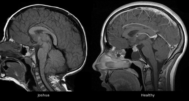
What Are The Causes Of Chiari Malformations
Primary CMs: these are the most common form caused mainly by structural defects in the brain and spinal cord during foetal development, as a result of genetic mutations or a maternal diet lackingcertain vitamins or nutrients.
Secondary CMs: caused later in life if spinal fluid is drained excessively from the lumbar or thoracic areas of the spine either due to traumatic injury, disease, or infection.
Understanding The Underlying Causes
The findings, published in the American Journal of Human Genetics, could lead to new ways to identify people at risk of developing Chiari 1 malformation before the most serious symptoms arise. It also sheds light on the development of the common but poorly understood condition. A lot of times people have recurrent headaches, but they don’t realise a Chiari malformation is the cause of their headaches. And even if they do, not everyone is willing to have brain surgery to fix it. Hence, there is a need for better treatments, and the first step to better treatments is a better understanding of the underlying causes.
London Spine Unit Harley Street Hospital
A Focus on High Quality Specialised Care
We are a specialist Private Hospital based on Harley Street, London UK The Harley Street Hospital, Day Surgery Hospital
We provide exclusive health services for individuals seeking Advanced medical, non-surgical or minimally invasive treatments. We are covered by All Insurance Companies apart from AXA PPP
Our Medical Director and Lead Spinal Surgeon Mr Mo Akmal MD is a world renowned Spine Specialist Consultant with over 20 years of experience. He and his team have developed revolutionary techniques to perform all types of Spinal Surgery as a Day Case procedure without traditional General Anaesthetic.
We are constantly improving our techniques for treatment and improving facilities for our patients.
Also Check: How Does Autism Affect Student Learning And Behavior
Some Chiarians Also Have
In the United States, scoliosis affects 2% of women and 0.5% of men in the general population. There are many causes of scoliosis, including the following: congenital spine deformities, genetic conditions, neuromuscular problems, limb length inequality, cerebral palsy, spina bifida, muscular dystrophy, spinal muscular atrophy and tumors. Over 80% of scoliosis cases, however, are idiopathic, which means that there is no known cause. Most idiopathic scoliosis cases are found in otherwise healthy people.The following are the three basic types of treatments for scoliosis: observation, orthopedic bracing, or surgery.AUTISM Autism and autism spectrum disorder are both general terms for a group of complex disorders of brain development. These disorders are characterized, in varying degrees, by difficulties in social interaction, verbal and nonverbal communication and repetitive behaviors.
FIBROMYALGIA SYNDROME AUTONOMIC DYSREFLEXIA
It can develop suddenly, and is a possible emergency situation. If not treated promptly and correctly, it may lead to seizures, stroke and even death.
Causes Of Chiari I Malformations

The exact cause of Chiari I malformations is unknown. It tends to be present from birth, but is normally only found in adulthood when symptoms develop or when an MRI scan is done.
Many cases are thought to be the result of part of the skull not being large enough for the brain.
Chiari I malformations can also develop in people with a tethered spinal cord, a build-up of fluid on the brain;, and some types of brain tumour.
Chiari malformations can sometimes run in families. Its possible that some children born with it may have inherited a faulty gene that caused problems with their skull development.
But;the risk of passing a Chiari malformation on to your child is very small. And remember: even if your children do inherit it,;they may not;experience symptoms.
Read Also: What Causes Autism Spectrum Disorder
E: Surgical Options For Specific Conditions Related To Cvj Malformations
A separate section was dedicated to the CVJ instability because of its still unclear relationship with CM1 and the still missing general agreement on its management, which is often different and even independent from CM1 . Particularly controversial is the use of fixation to manage CM1 patients without CVJ anomalies .
Table 6 Specific conditions related to CVJ malformations: surgical options
On these grounds, some questions were devoted to the definition and the diagnostic work-up of CVJ instability . The proposed definition includes also the clinical findings because instability is not ever easy to demonstrate radiologically. A good agreement was achieved about the need to perfect the diagnosis of CVJ instability with dynamic 3D CT scan and to carefully investigate the course of the vertebral artery and the volume and morphology of the occipital squama and cervical vertebrae prior to surgery. This appears particularly pertinent in children, where the anatomical conditions may vary significantly according to age. For the simple diagnosis, on the other hand, dynamic X-rays and MRI are enough .
Section 3Isolated/non-CM1 pediatric syringomyelia
How Is A Chiari Malformation Type I Treated
You may be treated by a neurologist or neurosurgeon. These are experts in brain and spinal cord problems. Treatment will depend on your symptoms, age, and general health. It will also depend on how severe the condition is.
-
With no symptoms. Your health may be watched closely. This may include frequent physical exams and MRI tests.
-
With symptoms. Your healthcare provider may prescribe medicines to reduce pain. Or he or she may choose surgery. This is done to relieve pressure on the brain or restore the flow of spinal fluid.
-
With few or no symptoms, but a syrinx. Your healthcare provider may suggest close monitoring of the defect with a special type of MRI called cine phase contrast. This test looks at the flow of spinal fluid. It also looks at areas where the fluid is blocked. You may need surgery, based on the MRI results or if symptoms get worse.
-
With signs of sleep apnea. You may need a sleep study if you have sleep apnea. Sleep apnea means that you stop and start breathing during sleep. A sleep study can also help your healthcare provider decide if you need other treatment.
Don’t Miss: Is Autism A Mental Illness Uk
Circulating Mirna Expression Profiling
A high-throughput expression analysis of 800 microRNAs in sera of 10 AC, 11 ACTS, 6 TS patients, and 8 NC subjects was computed by using nCounter NanoString technology.
We identified 9 miRNAs as significantly differentially expressed in sera from the different groups . More specifically: let-7b-5p upregulated in ACTS compared to TS patients; miR-21-5p upregulated in ACTS compared to AC patients and was downregulated in AC compared to TS patients; miR-23a-3p was upregulated in TS compared to NCs and was downregulated in AC compared to TS patients; miR-25-3p was upregulated in AC compared to TS patients and NCs and was downregulated in ACTS compared to AC patients; miR-93-5p was upregulated in AC compared to TS patients; miR-130a-3p was downregulated in ACTS and TS patients compared to NCs; miR-144-3p was downregulated in ACTS compared to AC patients and upregulated in AC compared to TS patients; miR-222-3p was upregulated in ACTS compared to NCs; miR-451a was upregulated in AC compared to TS patients and NCs, and in ACTS patients compared to NCs, with a FDR < 0.05 for each pairwise comparison. The relative expression of DE miRNAs is shown in Figure 1.
Table 3. Dysregulation of 9 miRNAs in different pairwise comparisons.
Clinical And Behavioral Characteristics Of Asd
Neurodevelopmental disorders typically manifest early in the developmental stage, most often before the child enters grade school; hence diagnosis and treatment of the neurodevelopmental disorders can be difficult. There are no specific tests that can predict developmental and cognitive deficits before early childhood in various neurodevelopmental disorders, including ASD.
Several characteristic features have been reported in ASD for early characterization of the disorders and commonly referred to as Red flags for the ASD. Various characteristic features have been reported at the various time point of the age, like no eye contact by 6 months of age, no response to name-calling, and no social referencing by the age of 10 months, no imitation and two meaningful words by 12 months of the age, no proto-declarative and proto-imperative pointing by 14 months of the age, and no joint attention by 18 months of age. Infants at risk for and later diagnosed with ASD showed a decline in eye fixation within the first 26 months of age. This pattern was not observed in typically developing infants. Children with autism showed lower rates of canonical babbling and fewer speech-like vocalizations across the age, i.e., 624 months of age than did typically developing peers.
You May Like: Is Autism Protected Under The Ada
Ethics Approval And Consent To Participate
All experiments were approved by the local ethical committee Comitato Etico Catania 1 prior to sample collection and in accordance with the Helsinki Declaration and its later amendments or comparable ethical standards. Written, informed consent was obtained from parents of all minor age participants .
Where Can I Get More Information
For more information on neurological disorders or research programs funded by the National Institute of Neurological Disorders and Stroke, contact the Institute’s Brain Resources and Information Network at:
Office of Neuroscience Communications and EngagementNational Institute of Neurological Disorders and StrokeNational Institutes of HealthBethesda, MD 20892
NINDS health-related material is provided for information purposes only and does not necessarily represent endorsement by or an official position of the National Institute of Neurological Disorders and Stroke or any other Federal agency. Advice on the treatment or care of an individual patient should be obtained through consultation with a physician who has examined that patient or is familiar with that patient’s medical history.
All NINDS-prepared information is in the public domain and may be freely copied. Credit to the NINDS or the NIH is appreciated.
Recommended Reading: Is Autism Genetic Or Hereditary
Volumetric Changes In Asd
Size of the brain is often measured by weight, sometimes by volume , and referred as cranial capacity. The changes in brain volume differ depending on several factors, such as age, environment, and body size. Mostly, the brain volume is measured in a cubic centimeter, and an average volume of a modern human brain is between 1300 and 1500cm3. Any deviation in brain volume results in structural changes in the brain tissue and might alter behavioral or functional patterns or vice versa. Studies on structural and volumetric changes in the ASD explored their relationship with behavioral changes. Findings from some of the recent studies in ASD using structural brain MRI are summarized below.
Infants with older ASD siblings are said to be at risk of developing ASD and other related neurodevelopmental issues, more specifically with male sibling,. A prospective neuroimaging study conducted by Hazlett et al. on infants at high risk for autism reported the hyperexpansion of cortical surface area between 6 and 12 months of age followed by an increase in brain volume between 12 and 24 months. Brain volume was associated with the emergence and severity of social deficits in ASD.
C: Diagnosis And Treatment Of The Main Causes Of Surgical Failure

Apart from the wrong indication to surgery and the occurrence of complications , some patient-related causes of failure have been reported in some series, such as comorbidities, unfavorable or complex anatomy, tonsils below C2, diameter of the spinal cord, and pediatric age . As far as the technique is concerned, the main cause of failure of the bony decompression alone is judged to be the too small bone opening/bone regrowth, especially at the level of the foramen magnum . In selected cases , 3D CT scan may be helpful in estimating the extent of the decompression and in planning a new operation to enlarge the bone opening.
As far as duraplasty is concerned, incomplete expansion duraplasty, arachnoid scarring, and wound-related complications are the main factors negatively affecting the outcome . Postoperative arachnoiditis is regarded as the most important cause of clinical and/or radiological recurrence. It can be addressed by a revision surgical procedure to perform a lysis of the adherences and/or to coagulate the tonsils or, in case of multiple recurrences, to stent the IV ventricle to maintain the patency of the obex . The role of the IV ventricle stenting is still under debate.
You May Like: Is Adhd Similar To Autism
Sample Collection And Processing
Peripheral blood samples from all participants were collected in the morning through a butterfly device, inserted into serum separation collection tubes with Clot activator and gel for serum separation . Collection tubes were treated according to current procedures for clinical samples. Tubes were rotated end-over-end at 20°C for 30 to separate serum from blood cells. Subsequently, they were centrifuged at 3,500 rpm at 4°C for 15 in a Beckman J-6M/E, supernatants were distributed into 1.5 ml RNase-free tubes, and finally stored at 80°C until analysis .
Key Points About Chiari Malformation Type I
-
With a Chiari malformation, the lower part of the brain dips down through a normal opening at the bottom of the skull.
-
There are several types of Chiari malformations. Type I is the most common type.
-
In most cases, the problem is present at birth . But it may not be found until a person is a teen or young adult.
-
You may not have symptoms. If symptoms occur, the most common ones are headaches or pain in the back of the head or neck. The headaches and pain are made worse by coughing, laughing, or sneezing.
-
You may also have a pocket of spinal fluid in the spinal cord or brain stem. This is called a syrinx.
-
Imaging tests are done to detect a Chiari malformation type I. You may have an MRI or a CT scan.
Read Also: Is Autism A Social Issue
What Does An Upper Cervical Chiropractor Understanding How Blair Upper Cervical Can Help Those That Suffer From Chiari Malformation
Blair Upper Cervical Chiropractors are well-trained specialty doctors who focus ALL of their attention on the relationship of the skull, atlas, axis, and their relationship to the brainstem and associated spinal nerves. ;The Blair Upper Cervical Doctor runs a battery of tests to locate spinal cord pressure and interference resulting from spinal misalignment. ;
Once the patient is determined in having spinal misalignment precise imaging is taken to using cone beam computed tomography or digital x-ray to visualize the joints and how they fit. Each joint in the upper cervical spine fits as a mirror image with the other. If spinal misalignment exists by viewing each joint the doctor can determine exactly how the vertebrae have misaligned from normal and determine the angulation of the joint.;
This information, which is unique to each individual, is then used in making a precise spinal correction without using any twisting, popping, or pulling. ;The goal of the care is to stabilize the biomechanics of the upper cervical spine, so the central nervous system can function without structural interference. The correction of the upper cervical spine allows the central nervous system to function more normally.;
To schedule you can call 310 324-6172 for our Carson office or 213 399-7772 for our Los Angeles office.;
If you are outside of the Los Angeles area you can call our office and we would be happy to find you an upper cervical doctor in your location. ; ;;
References:
Leave a comment
Banking On Brains For Clues To Autism
New initiatives aim to increase brain donations for autism research and maximize what scientists can learn from these precious specimens.
by Katie Moisse;/;1 November 2017
Leslie Bolen was there for every one of her son Michaels medical tests. Hed had more than his fair share in his 14 years, but this last one was almost too much for his mother to bear.
That test was on a Saturday night in April 2016. Eight days earlier, Michael had had a seizure at the residential facility where he lived. He had autism, and hed had seizures almost every week since he was 8 years old, but never one this bad. He stopped breathing, and his heart stopped beating. Medics managed to restart his heart with a shot of adrenaline, but his brain never recovered.
Michael spent the following week on life support. His parents rarely left his side, ducking out of the hospital only three times for clean clothes and to spend time with their then-13-year-old daughter, Rachel. Sitting in a chair beside Michaels bed, Bolen played back events from the previous Friday in her mind: the frantic call from Michaels caseworker, the seemingly endless 10-minute drive to the hospital, the look on her husband Chads face when he arrived at the hospital.
Unthinkable gift: Leslie Bolen donated her sons brain with hopes it would eventually help others with autism.
Read Also: What Is The Definition Of Autism
Enrichment And Correlation Analysis Of Serum Mirnas In Comorbidity Between Arnold
- 1Section of Biology and Genetics Giovanni Sichel, Department of Biomedical and Biotechnological Sciences, University of Catania, Catania, Italy
- 2Section of Child and Adolescent Psychiatry, Department of Clinical and Experimental Medicine, University of Catania, Catania, Italy
- 3Radiology Unit 1, Department of Medical Surgical Sciences and Advanced Technologies, University Hospital Policlinico-Vittorio Emanuele, University of Catania, Catania, Italy
- 4Oasi Research InstituteIRCCS, Troina, Italy
Neuroimaging Studies In Asd
Brain development throughout the life span is a complex and dynamic process that should be considered early infancy, even prenatally. Recently, the rapid development of noninvasive brain imaging technology, a new generation of imaging technique such as MRI, holds great promise for its ability to reveal structural and functional brain alterations during development in infants, children, and adolescents . Given its noninvasive nature, MRI could be used in clinical practice as part of the comprehensive clinical assessment of ASD patients to exclude brain alterations. As the relationship between the postnatal CNS structural development and functional capacities of children from birth to adolescence gradually unfolds, the potential for identifying early delays in cognitive development mounting effective treatment and prevention strategies increases. Thus, it allows pre-emptive interventional resources and biological-based therapies for targeted intervention to reduce the frequent burden of intellectual disabilities in the ASD population. Although studies using structural and functional MRI have highlighted alterations in the neuroanatomy of ASD in the past few decades,,, a univocal, reliable, and consistent pattern of alterations is yet to be identified. Also, there is not enough information available on imaging perspective to clearly understand the underlying pathophysiology of ASD at a younger age, i.e., below 6 years of age.
You May Like: How Do You Get Diagnosed With Autism