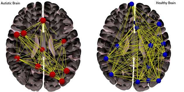
Brain Imaging For Autism Diagnosis
Physics is the science of the universal: finding common rules that govern the behaviors of disparate systems. But medical physicists have a different approach, as many diseases call for customizedrather than blankettreatment plans. That is where a new technique for characterizing structures in the brain might come in handy, hopes Shannon Brindle, a final-year undergraduate physics major working in the lab of Rasha Makkia at the University of Mary Washington, Virginia. For example, she envisions that advanced brain imaging analysis techniques could help clinicians verifyat a younger age than currently possiblewhether a child has an autism spectrum disorder, pinpoint its severity, and then better tailor interventions to that childs individual needs. Brindle discussed her findings in a talk at this years March Meeting of the American Physical Society.
One in 54 children in the US receives an autism diagnosis each year, 5 times more than the number of children who develop cancer. But no blood or imaging tests exist for the disorder. Rather, the disorder, which develops in early childhood, is diagnosed by studying a childs ability to communicate and interact with other people. That means it can take time for a family to receive a formal diagnosis, which can delay treatment and leave parents fretting for answers.
Katherine Wright
Katherine Wright is the Deputy Editor of Physics.
Autism Is Detectable In Brain Scans Long Before Symptoms Appear New Study Says
Researchers at the University of North Carolina used brain scans to predict with 80% accuracy whether certain children would develop autism, meaning such scans could enable early treatment of the disorder long before symptoms are evident.
Until recently, the average age of diagnosis for autism was about four years, NBC News reported, but the new research suggests autism may be detectable within a child’s first year. And that’s important, said Dr. Joseph Piven, who lead authored a resulting study.
“If we can target interventions before autism appears and before the brain changes appear, during a time when the brain is highly malleable or plastic, we can have a bigger impact on the outcome,” Piven, who directs the university’s Carolina Institute for Developmental Disabilities, told NBC News.
Autism affects about 1 in 100 people in the general population, according to the journal Nature, which published the small study. But Infants with an older sibling who has autism have about a 1 in 5 chance of developing autism spectrum disorder.
The study involved MRI scans on 106 infants with older siblings who had autism and 42 infants whose families had no history of the disorder, Nature reported. The scans, taken at six months, one year and two years, revealed that children later diagnosed with autism showed key brain growth during their first year, the university said.
White Matter: Connecting The Clinical Dots
The second study, also published in Biological Psychiatry, linked changes in the brains white matter growth with autism traits in some children.
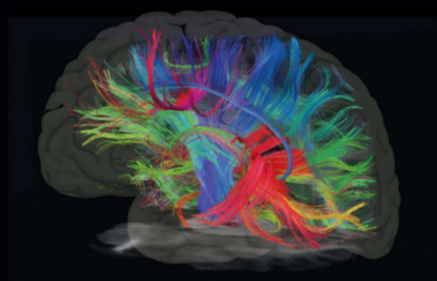
The researchers used a type of MRI scan called diffusion-weighted imaging, which allowed them to look at white matter regions, or tracts, in the brain. White matter provides the structural connections in the brain, allowing different regions to communicate with each other.
The study included 125 children with autism and 69 typically developing children who served as controls, between the ages of 2.5 and 7.
The researchers found that the development of the white matter tracts in the brain was linked to changes in autism symptom severity. They observed slower development in children whose symptom severity increased over time, and faster development in those with decreased severity over time.
From a biological standpoint, this emphasizes the role of white matter development in autism and autism symptoms, said Derek Sayre Andrews, postdoctoral scholar at the MIND Institute and lead author on the paper. We hope that in the future, measurements like this can identify children who would benefit from more intensive intervention and serve as a marker to determine the effectiveness of an intervention for a particular child, he said.
Recommended Reading: Stage 3 Autism
Brain Scans Show Early Signs Of Autism Spectrum Disorder
Posted on by Dr. Francis Collins
Getty Images
For children with autism spectrum disorder , early diagnosis is critical to allow for possible interventions at a time when the brain is most amenable to change. But thats been tough to implement for a simple reason: the symptoms of ASD, such as communication difficulties, social deficits, and repetitive behaviors, often do not show up until a child turns 2 or even 3 years old.
Now, an NIH-funded research team has news that may pave the way for earlier detection of ASD. The key is to shift the diagnostic focus from how kids act to how their brains grow. In their brain imaging study, the researchers found that, compared to other children, youngsters with ASD showed unusually rapid brain growth from infancy to age 2. In fact, the growth differences were already evident by their first birthdays, well before autistic behaviors typically emerge.
Autism spectrum disorder includes a range of developmental conditions, such as autism and Asperger syndrome, that are characterized by challenges in social skills and communication. Scientists have long known that teens and adults with ASD have unusually large brain volumes. Researchers, including Heather Hazlett and Joseph Piven of the University of North Carolina, Chapel Hill, found more than a decade ago that those differences in brain size emerge by about age 2 . However, no one had ever visually tracked those developmental differences.
References:
Differences In Autistic Brains

That’s because the brains of people with autism tend to have structural differences in parts of the frontal and parietal lobesareas of the brain involved in behavior and language.
The scan needs more testing before it’s put to widespread use. But if it pans out, the scan could help doctors diagnose autism more quickly and more accurately. And that, in turn, could lead to earlier and more effective therapies.
You May Like: Autism Awareness Color Meanings
Early Brain Imaging In Infants May Help Predict Autism
JAMA. 2017 318:1211-1212. doi:10.1001/jama.2017.13706
Infants at high familial risk of autism spectrum disorder do not typically exhibit symptoms in their first year of life, but new research indicates that magnetic resonance imaging may reveal signs of the disorder during this presymptomatic period. The findings point to a noninvasive method to detect autism at its earliest stages, when interventions may provide the most benefits.
Prospective neuroimaging findings suggest increased cerebral cortical growth between 6 and 12 months of age may predict autism diagnosis at 24 months of age.
An estimated 1 in 5 infants who have an older sibling with ASD develop the condition, compared with approximately 1 in 100 in the general population. Because the defining features of ASD tend to emerge over the latter part of the first year and into the second year, a diagnosis is not typically made until 24 months of age and beyond.
Two prospective neuroimaging studies published earlier this year in Nature and Science Translational Medicinetrained machine learning algorithms using longitudinal MRI data from infants at high familial risk of ASD to predict autism diagnosis. The first study published in Nature compared behavioral changes and brain structural changes in 106 infants at high familial risk of ASD and 42 low-risk infants at 6, 12, and 24 months of age.
How Mris Can Be Used To Diagnose Autism
Though we have made progress in recognizing what does not cause autism, little is currently known about what does cause most cases of autism. It is known that there is no single cause of the condition. In general, it is accepted that autism is related to structural differences in the brain, though not much is understood about the specific differences. Research suggests that autism may be caused by a combination of genetic or nongenetic, or environmental, influencers. And, while certain environmental factors are thought to increase risk, this is certainly not the same thing as directly causing autism. Its well known that all of these factors affect brain development and communication between different regions of the brain, but the specific mechanisms are not as well-understood.
Also Check: Life Expectancy Of Someone With Autism
Specialized Mri Shows Autism Changes Brains White Matter Significantly
Researchers at Yale University analyzing specialized MRI exams found significant changes in the microstructure of the brains white matter in adolescents and young adults with autism spectrum disorder compared to a control group, according to research being presented next week at the annual meeting of the Radiological Society of North America . The changes were most pronounced in the region that facilitates communication between the two hemispheres of the brain.
One in 68 children in the U.S. is affected by ASD, but high variety in symptom manifestation and severity make it hard to recognize the condition early and monitor treatment response, said Clara Weber, postgraduate research fellow at Yale University School of Medicine. We aim to find neuroimaging biomarkers that can potentially facilitate diagnosis and therapy planning.
Researchers reviewed diffusion tensor imaging brain scans from a large dataset of patients between the age of six months and 50 years. DTI is an MRI technique that measures connectivity in the brain by detecting how water moves along its white matter tracts. Water molecules diffuse differently through the brain, depending on the integrity, architecture, and presence of barriers in tissue.
Significant alterations in the brains white matter in adolescents with autism spectrum disorder . Credit: RSNA and researcher, Clara Weber
A Personalized Autism Diagnosis Cad System Using A Fusion Of Structural Mri And Resting
- 1Bioimaging Lab, Bioengineering Department, University of Louisville, Louisville, KY, United States
- 2Department of Electrical and Computer Engineering, Abu Dhabi University, Abu Dhabi, United Arab Emirates
- 3Department of Biomedical Sciences, University of South Carolina, Greenville, SC, United States
- 4Computer Engineering and Computer Science Department, University of Louisville, Louisville, KY, United States
- 5Bioengineering Department, University of Louisville, Louisville, KY, United States
- 6Department of Neurology, University of Louisville, Louisville, KY, United States
Don’t Miss: Pivotal Behavior Examples
Uc Davis Mind Institute Researchers Tracked Brain Changes In Children Over Many Years Using Mri Scans
This video is best viewed in Chrome or Firefox.
Two groundbreaking studies at the UC Davis MIND Institute provide clues about possible types of autism linked to brain structure, including size and white matter growth.
The research is based on brain scans taken over many years as part of the Autism Phenome Project and Girls with Autism, Imaging of Neurodevelopment studies. It shows the value of longitudinal studies that follow the same children from diagnosis into adolescence.
The researchers tracked brain growth and structure in hundreds of children from age 3 to age 12
There is no other single site data set like ours anywhere, said Christine Wu Nordahl, associate professor in the Department of Psychiatry and Behavioral Sciences, MIND Institute faculty member and co-senior author on both papers. In one of the studies we have over 1,000 MRI scans from 400 kids, which is unheard of. Its been 15 years of work to get here.
What Is The Role Of Mri In The Workup Of Autism Spectrum Disorder
Magnetic resonance imaging studies in patients with autism yield inconsistent results. However, typical findings include enlargement of the total brain, the total brain tissue, and the lateral and fourth ventricles, along with reductions in the size of the midbrain, the medulla oblongata, the cerebellar hemispheres, and vermal lobules VI and VII. Although vermal hypoplasia is found in some individuals with autism, vermal hyperplasia is identified in others.
The volume of the gray matter is bilaterally decreased in the amygdala, the precuneus, and the hippocampus of people with autism spectrum disorder. Adolescents with autism spectrum disorder have shown greater decreases in the volume of the gray matter of the right precuneus than have adults. The volume of the gray matter in the middle-inferior frontal gyrus has been found to be slightly increased in people with autism spectrum disorder.
Imaging studies in patients with autistic disorder who exhibit head banging may show enlargement of the diploic space in the parietal and occipital bones, with loss of gray matter adjacent to the bony changes. These findings resemble those of posttraumatic encephalopathy in athletes in contact sports and professional boxers .
References
Lord C, Elsabbagh M, Baird G, Veenstra-Vanderweele J. Autism spectrum disorder.Lancet. 2018 Aug 11. 392 :508-520. .
Courchesne E, Carper R, Akshoomoff N. Evidence of brain overgrowth in the first year of life in autism. JAMA. 2003 Jul 16. 290:337-44. .
Recommended Reading: Life Expectancy Of Autistic Person
Can Brain Scans Help Personalize Autism Therapies And Supports
This Autism Speaks research fellow has developed a test that helps predict who will respond best to certain behavioral therapies and coaching programs
An Autism Speaks Meixner Postdoctoral Fellowship in Translational Research supported the research Dr. Yang describes here.
Im glad to tell you about the promising findings of the research supported by my Autism Speaks Meixner Fellowship. My team is developing and testing methods that predict how well someone with autism will respond to certain behavioral therapies and social coaching programs.
The goal is to provide guidance to families and individuals affected by autism so they can make informed choices for treatment and support. Its also in line with the growing appreciation that each person with autism is unique and does best with a personalized intervention program.
Knowing who will benefit most from an intervention and at what point in their lives will also allow us to adjust treatment approaches to maximize benefit, avoid excessive costs and in some cases flag when a different or more-intensive program is more likely to produce the desired benefits.
Heterogeneity And Other Diagnostic Challenges

Autism is inherently heterogeneous and the condition involves plenty of uncertainties that could complicate diagnosis. Furthermore, it occurs on a wide spectrumas the well-known saying goes: If youve met one individual with autism, youve met one individual with autism. .
As diagnosis for the condition involves behavioral evaluations, a further complication involves masking or camouflaging of symptoms found especially among females on the spectrum. All these uncertainties and challenges fuel the research for brain biomarkers for neurodevelopmental disorders.
Establishing neuroimaging biomarkers of autism using MRI may be a crucial step for accurate diagnosis and better, tailored treatment. This may be especially important early on in a child on the spectrums life when appropriate intervention may have the greatest effect .
Recommended Reading: Autism And Stuttering
Why Choose Amen Clinics For Autism Spectrum Disorder Treatment
In addition to understanding the ASD brain patterns, there are other ways the brain SPECT imaging used at Amen Clinics can help. Children with autism often struggle with other mental health conditions, such as ADD/ADHD, depression, and anxiety. According to a growing body of research, over 70% of children with ASD have at least one additional co-existing medical or psychiatric condition, and over 40% have two or more such conditions. SPECT brain imaging can help show if these conditions are present in addition to ASD so that treatment can be targeted to address all of the issues you or your child have. Be aware that the sooner a child with autism gets help, the more effective treatment will be. Early intervention can help with your childs overall development and decrease symptoms as they grow up. Its important to realize that adults can also benefit from treatment at any age.
In Which Way Does The Autistic Brain Look Different
A differently wired brain is often mentioned when referring to autism. Increasingly, studies are showing that there are actual differences in the autistic brain using MRI, researchers identified structural abnormalities in the brains of individuals with one of the most common genetic causes of autism.
The science behind some of these brain differences may be complicated, but it seems to come down to abnormalities that many people with ASD have, at the 16th chromosome called 16p11.2. At this site, deletion or duplication of a piece of a chromosome may result in one of the most common genetic causes of autism.
The study revealed some interesting brain differences in deletion and duplication carriers in comparison to non-carriers:
- In comparison to non-carriers and control groups, the fiber bundle connecting the left and rights hemispheres of the brain was differently shaped and thicker in the deletion carriers and thinner in the duplication carriers
- Brain overgrowth was apparent in the deletion carriers
- The duplication carriers, on the other hand, displayed features of brain undergrowth
In this study differences in brain structure, visible on brain images, corresponded to behavioral and cognitive deficits. These results may have implications for future diagnosis of autism, if structural brain abnormalities are easily identifiable using brain scans, autism could be identified earlier with more accuracy.
Recommended Reading: Autism Spectrum Symbol
Overly Persistent Brain Connections
First, the researchers conducted functional MRI scans on 90 male participants, of which 52 had a diagnosis of autism and 38 did not. The participants with autism were aged between 19 and 34, while the rest of the volunteers who acted as the control group had ages ranging between 20 and 34.
Then, to confirm the initial findings, the specialists compared their data with that collected from a further 1,402 people who participated in the Autism Brain Imaging Data Exchange study. Of these, 579 participants had autism. The remaining 823 participants did not have autism and acted as the control group.
Dr. Anderson and team used a novel fMRI method to explore brain activity in the participants on the current study. More specifically, they looked at the duration of connections established across brain regions.
We dont have good methods for looking at the brain on these timescales. Its been a blind spot because it falls in between typical MRI and studies, explains Dr. Anderson.
Thanks to the fMRI scans, the researchers were able to confirm that in the brains of people with autism, connections persist for more extended periods than they do in the brains of neurotypical individuals. In other words, in autism, the brain finds it harder to switch between processes.